


Order NO. D290056
兔抗AKT1/AKT2/AKT3单克隆抗体
| 英文名 | Anti-AKT1/AKT2/AKT3 rabbit monoclonal antibody | ||
| 别名 | AKT serine/threonine kinase 1/2/3; AKT; PKB; RAC; PRKBA; PKB-ALPHA; RAC-ALPHA; PKBB; PRKBB; HIHGHH; PKBBETA; RAC-BETA; MPPH; PKBG; M | ||
| 相关类别 | 一抗 | 储存 | 冷冻(-20℃) |
| 宿主 | Rabbit | 反应种属 | Human, Mouse, Rat, Hamster |
| 标记物 | Unconjugate | 克隆类型 | rabbit monoclonal |
| 抗原 | AKT1/AKT2/AKT3 | ||

|
|||
| 编 号 | 包装 | 库存 | 目录价(¥) | 您的价格(¥) | 数 量 |
| D290056-0050 | 50 ul | 预计5日内发货 |
|
|
|
| D290056-0100 | 100 ul | 预计5日内发货 |
|
|
|
属性
| Background: | The AKT1 gene encodes one of the three members of the human AKT serine-threonine protein kinase family which are often referred to as protein kinase B alpha, beta, and gamma. These highly similar AKT proteins all have an N-terminal pleckstrin homology domain, a serine/threonine-specific kinase domain and a C-terminal regulatory domain. These proteins are phosphorylated by phosphoinositide 3-kinase (PI3K). AKT/PI3K forms a key component of many signalling pathways that involve the binding of membrane-bound ligands such as receptor tyrosine kinases, G-protein coupled receptors, and integrin-linked kinase. These AKT proteins therefore regulate a wide variety of cellular functions including cell proliferation, survival, metabolism, and angiogenesis in both normal and malignant cells. AKT proteins are recruited to the cell membrane by phosphatidylinositol 3,4,5-trisphosphate (PIP3) after phosphorylation of phosphatidylinositol 4,5-bisphosphate (PIP2) by PI3K. Subsequent phosphorylation of both threonine residue 308 and serine residue 473 is required for full activation of the AKT1 protein encoded by this gene. Phosphorylation of additional residues also occurs, for example, in response to insulin growth factor-1 and epidermal growth factor. Protein phosphatases act as negative regulators of AKT proteins by dephosphorylating AKT or PIP3. The PI3K/AKT signalling pathway is crucial for tumor cell survival. Survival factors can suppress apoptosis in a transcription-independent manner by activating AKT1 which then phosphorylates and inactivates components of the apoptotic machinery. AKT proteins also participate in the mammalian target of rapamycin (mTOR) signalling pathway which controls the assembly of the eukaryotic translation initiation factor 4F (eIF4E) complex and this pathway, in addition to responding to extracellular signals from growth factors and cytokines, is disregulated in many cancers. Mutations in this gene are associated with multiple types of cancer and excessive tissue growth including Proteus syndrome and Cowden syndrome 6, and breast, colorectal, and ovarian cancers. Multiple alternatively spliced transcript variants have been found for this gene. The AKT2 gene is a putative oncogene encoding a protein belonging to a subfamily of serine/threonine kinases containing SH2-like (Src homology 2-like) domains, which is involved in signaling pathways. The gene serves as an oncogene in the tumorigenesis of cancer cells For example, its overexpression contributes to the malignant phenotype of a subset of human ductal pancreatic cancers. The encoded protein is a general protein kinase capable of phophorylating several known proteins, and has also been implicated in insulin signaling. The protein encoded by the AKT3 gene is a member of the AKT, also called PKB, serine/threonine protein kinase family. AKT kinases are known to be regulators of cell signaling in response to insulin and growth factors. They are involved in a wide variety of biological processes including cell proliferation, differentiation, apoptosis, tumorigenesis, as well as glycogen synthesis and glucose uptake. This kinase has been shown to be stimulated by platelet-derived growth factor (PDGF), insulin, and insulin-like growth factor 1 (IGF1). Alternatively splice transcript variants encoding distinct isoforms have been described. |
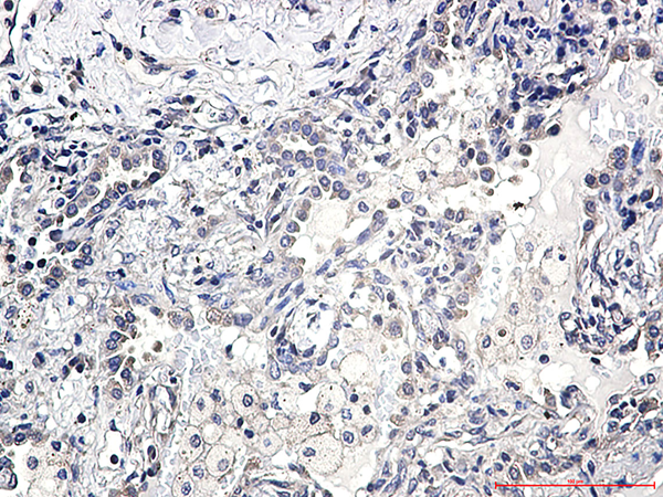
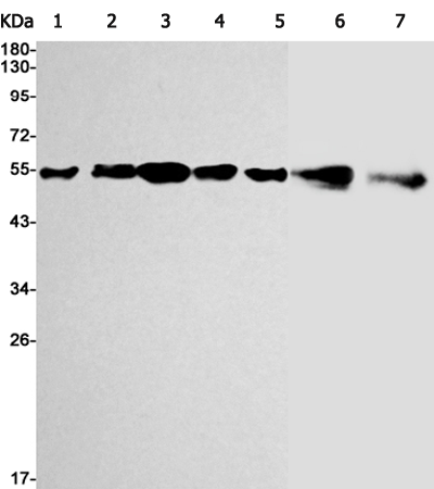
| Applications: | WB, IHC |
| Name of antibody: | AKT1/AKT2/AKT3 |
| Immunogen: | Fusion protein of human AKT1/AKT2/AKT3 |
| Full name: | AKT serine/threonine kinase 1/2/3 |
| Synonyms: | AKT; PKB; RAC; PRKBA; PKB-ALPHA; RAC-ALPHA; PKBB; PRKBB; HIHGHH; PKBBETA; RAC-BETA; MPPH; PKBG; MPPH2; PRKBG; STK-2; PKB-GAMMA; RAC-gamma; RAC-PK-gamma |
| SwissProt: | P31749/P31751/Q9Y243 |
| IHC positive control: | Human lung cancer |
| IHC Recommend dilution: | 50-100 |
| WB Predicted band size: | 56 kDa |
| WB Positive control: | Jurkat, Rat brain, C6, CHO-K1, Hela, 3T3, Mouse lung lysates |
| WB Recommended dilution: | 500-2000 |
相关文档
|
|
||||
Sample Application

